

Causes and Treatments of Tear and Saliva Staining in Dogs and Cats
The reddish-brown streaks beneath a Maltese’s eyes or along a Bichon’s muzzle are so familiar they can start to seem like a breed characteristic rather than a medical question worth asking. Tear staining, and the similar discoloration that comes from chronic licking, are caused by porphyrins, pigment compounds found in tears and saliva that oxidize and stain light-colored fur. But why the excess moisture is there in the first place is the more important question: shallow eye sockets, hair that contacts the cornea, blocked tear ducts, dietary factors, chronic low-grade infection, and dental disease are all contributors that vary from pet to pet. Addressing the staining without addressing the source is cosmetic work, not medical care.
Spring Branch Veterinary Hospital in Spring Branch, TX, has longer appointment times and a Fear Free certified team that takes the time with every patient needed to actually investigate what is driving a concern rather than just manage its appearance. Contact us to have a tear or saliva staining concern properly evaluated.
Why Do Pets Get Reddish-Brown Staining on Their Fur?
If you have noticed the rust-colored tracks under a dog’s eyes or the discolored fur between a cat’s toes, you have seen porphyrins at work. Porphyrins are natural compounds found in tears, saliva, and urine. When exposed to air and light over time, they oxidize and darken, staining whatever surface they land on. Light-colored coats simply make the problem more visible; darker pets may have the same underlying issues without the obvious visual evidence.
Tear stains form when tears overflow onto the face instead of draining normally through the tear ducts. The moisture creates a warm, damp environment against the skin where yeast and bacteria thrive, which is why stained areas often have a musty odor and can develop secondary skin irritation over time. Saliva staining on paws, legs, and muzzles follows the same logic: chronic dampness from licking produces the same porphyrin oxidation, with the same risk of secondary infection.
Brachycephalic breeds are particularly prone to facial tear staining because their flattened facial structure positions the eyes more prominently, reduces the space for normal tear drainage, and often brings facial skin folds into contact with the eye. Breeds like Pugs, Bulldogs, Shih Tzus, Persians, and Himalayans deal with this structurally, though any dog or cat with heavy jowls, prominent eyes, or long facial hair around the eyes can develop significant staining.
The key point is that staining is a symptom, not the problem itself. Our team evaluates the full picture during wellness exams, checking eye health, skin condition, dental health, and overall wellbeing to identify contributing causes.
What Eye Problems Cause Excessive Tearing?
Structural Issues That Block Normal Tear Drainage
Tears normally drain through small openings called puncta located near the inner corner of each eyelid, flowing through small canals into the nasal passages. When that drainage system is disrupted, tears have nowhere to go but down the face.
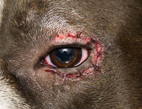
Exophthalmos, or abnormal protrusion of the eye, is common in flat-faced breeds and changes the geometry of how tears drain. Entropion is a condition where the eyelid rolls inward, bringing lid tissue into contact with the eye surface and causing chronic irritation and tear production. Eyelash disorders such as distichiasis or ectopic cilia cause constant mechanical irritation that stimulates tearing. Nasolacrimal duct obstruction, where the drainage canal itself becomes blocked, means tears have no path to drain and simply overflow. Some structural issues resolve with medical management, others improve with grooming changes, and some require surgical correction. Our team evaluates eyelids, lashes, and tear drainage as part of every staining workup.
Irritation, Infection, and Other Causes of Increased Tear Production
Eyes produce more tears when they hurt or are inflamed. Corneal ulcers, which are surface scratches or erosions on the clear front of the eye, are painful and trigger significant tear production. They are diagnosed using a fluorescent dye test during examination and require specific treatment to heal properly. Conjunctivitis, inflammation of the tissue lining the eyelids, causes redness, discharge, and increased tearing that can lead to staining.
Dry eye, or keratoconjunctivitis sicca, is a condition where the eye does not produce enough of the aqueous portion of the tear film. Counterintuitively, this can trigger reflex tearing from irritation, while the underlying quality of tear production is insufficient. Glaucoma, elevated pressure within the eye, similarly causes reflex tearing alongside other serious signs. Environmental irritants common in the Texas Hill Country, including cedar and oak pollen, dust, and smoke, can drive seasonal increases in tear production and staining.
A thorough eye examination evaluates the full eye surface, eyelid position, tear production, and the presence of ulcers or foreign material. Warning signs that warrant prompt evaluation: squinting, pawing at the face, visible redness or cloudiness, thick or colored discharge, or any sudden change in the appearance of the eye.
Could Allergies Be Causing the Staining?
Environmental and Seasonal Triggers
Allergies are one of the most common drivers of both tear and saliva staining, and in central Texas, the allergen burden is significant for a large portion of the year. Mountain cedar in winter, oak and grass pollen in spring, and mold spores after rain all affect sensitized pets. Dogs and cats with atopic dermatitis, the veterinary term for environmental allergies, experience skin and eye inflammation that drives tearing, facial rubbing, and the compulsive licking and chewing that produces saliva staining on paws and legs.
Patterns are useful diagnostic information: staining that worsens in spring or fall, paw licking that spikes after walks outdoors, or facial rubbing that began when a new cleaning product was introduced all point toward environmental triggers. Comprehensive allergy management addresses the inflammatory cause rather than managing symptoms alone, and may include anti-itch medications, topical treatments, and in appropriate cases, immunotherapy to reduce long-term sensitivity.
Food Sensitivities and Diet-Related Triggers
Some pets react to specific proteins or other ingredients in their diet, producing chronic skin, eye, and GI inflammation year-round without seasonal patterns. An elimination diet trial using a novel protein or hydrolyzed diet is the most reliable way to investigate this.
A proper diet trial for skin and eye symptoms runs eight to twelve weeks with no treats, table scraps, flavored supplements, or pill pockets made from other proteins. Strict adherence is what makes the result meaningful. If staining and other symptoms improve significantly on the trial food and return when the original diet is reintroduced, dietary sensitivity is confirmed and long-term management focuses on consistent diet choice. Our team can guide which trial diet makes sense based on a specific patient’s food history.
Other Health Conditions That Drive Staining
Excess licking that causes saliva staining has a broad range of possible drivers, and the source is not always the area being licked. Dental disease is one of the most commonly overlooked contributors to facial staining. Infected teeth and inflamed gums increase drooling and cause pets to paw at their faces, generating persistent moisture and staining around the muzzle. Our dentistry services include professional cleaning and oral health evaluation that address this directly.
Arthritis and chronic pain cause pets to lick painful joints, often producing brown staining on legs, hips, and feet that gets attributed to allergies when the real driver is discomfort. Laser therapy and geriatric care at our hospital offer effective options for pets managing chronic pain that is contributing to compulsive licking.
Other contributors include anxiety and compulsive grooming behaviors, and parasites. Flea allergy dermatitis drives widespread licking and chewing; consistent parasite prevention reduces several of these triggers and is part of the foundation of managing any pet with chronic licking and staining.
How Is the Cause of Staining Diagnosed?
The diagnostic process at Spring Branch Veterinary Hospital starts with a thorough history. When did staining begin? Has it changed recently? Are there other symptoms like scratching, head shaking, or changes in behavior? What does the pet eat, and has that changed? Are there seasonal patterns? These details guide the physical examination and determine which additional tests will be most informative.
During the exam, our team evaluates eyelid position and health, lash placement, tear production using a Schirmer tear test, the eye surface using fluorescent dye to identify ulcers, and the drainage system. The skin around stained areas is assessed for secondary yeast or bacterial overgrowth.
Skin cytology examines cells collected from the skin surface under a microscope and identifies whether infection is present and what organisms are involved. Dental health, joint comfort, and potential behavioral contributors are all evaluated. Radiology and in-house lab capabilities allow quick results for basic testing, with referral lab options available for more specialized panels.
Managing Staining at Home
Daily Cleaning Habits That Actually Help
Consistent daily cleaning prevents stains from setting into the fur and keeps secondary yeast and bacteria in check. Eye cleaning technique matters: use a veterinarian-approved wipe or saline-moistened cotton, work from the inner corner of the eye outward, and use a fresh pad for each eye to avoid transferring material from one side to the other. Vet Classics Tear Stain Finger Wipes are designed specifically for this task.
Daily home management includes the following practical steps:
- Keep the area around the eyes and muzzle dry throughout the day
- Keep fur trimmed so it does not wick moisture against the skin
- Brushing and grooming around eyes regularly helps prevent buildup before stains set
- For paw staining, wipe feet after outdoor time and trim the fur between pads
- Watch for signs of secondary skin problems developing
Products to Avoid
Not all tear stain products marketed to pet owners are safe. Unapproved tear stain products that contain antibiotics are sold without FDA approval. Long-term antibiotic exposure through these products contributes to antibiotic resistance and may cause permanent tooth discoloration. Hydrogen peroxide and bleaching products can cause chemical burns to delicate eye tissue and skin.
Stick to veterinarian-recommended cleaning products and get guidance before trying anything sold online with impressive before-and-after photos.
Preventing Secondary Yeast and Bacterial Infections
Keeping stained areas dry is the most effective prevention for secondary infection. Practical steps include using elevated food and water bowls if your pet’s beard stays wet after drinking, switching from plastic bowls to stainless steel or ceramic options that do not harbor bacteria in surface scratches, and washing bowls daily.
Signs of secondary infection that warrant a veterinary visit: foul odor from stained areas, redness or swelling of the skin beneath the fur, tenderness when the area is touched, or your pet scratching or rubbing the stained area more than usual.
When Should You Call the Clinic?
Staining that develops gradually over months is different from staining that appears suddenly or worsens quickly. Gradual onset often reflects slowly developing conditions; sudden change can indicate a new, acute problem.
Contact us or request an appointment if your pet is showing:
- Squinting, pawing at the face, or sensitivity to light
- Yellow, green, or white eye discharge rather than clear tearing
- Redness, visible swelling, or cloudiness of the eye
- A foul odor from stained areas or visible skin irritation beneath the fur
- Sores, hot spots, or bleeding where your pet has been licking
- One-sided staining that appeared suddenly, which can indicate a localized problem on that side
Our Fear Free certified approach makes these visits less stressful for anxious pets, and longer appointment times mean our team can address concerns thoroughly.

Frequently Asked Questions About Tear and Saliva Staining
Do certain breeds stain more than others?
Yes. Flat-faced breeds with prominent eyes and reduced tear drainage produce more staining as a structural reality. Pets with white or light-colored coats show staining more visibly, though any pet can be affected. Heavy-jowled breeds that drool more also tend to have more saliva staining around the muzzle.
Are over-the-counter stain remover products safe?
Not all of them. Products containing tylosin or other antibiotics are not FDA-approved for use in dogs or cats and carry real risks including antibiotic resistance and permanent tooth discoloration. Stick to products your veterinary team recommends.
Clear Eyes and Comfortable Skin: A Team Effort
Tear and saliva staining is rarely just a grooming problem. Behind the brown streaks and discolored paws is a reason: anatomy, infection, allergy, pain, dental disease, or diet. Finding that reason and addressing it is what produces lasting improvement rather than temporary cosmetic results.
Spring Branch Veterinary Hospital takes time with every patient to do exactly that. Whether the cause turns out to be a structural eyelid issue, an environmental allergy to Hill Country cedar, or a dental abscess driving excess drooling, our team is equipped to identify it and help you manage it. Contact us or request an appointment to have your pet’s staining properly evaluated and start treating the cause, not just the color.


Leave A Comment